
21 Jul Detailed view of viral replication machinery lends new insights into infection
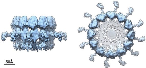
Cryoelectron microscope tomography imaging reveals high-resolution side and top views of the viral RNA replication “crown” complex structure. COURTESY OF PAUL AHLQUIST
The coronavirus that causes COVID-19, SARS-CoV-2, is known as a positive-strand RNA virus because of the way it stores and makes copies of its genetic material. Many other important pathogens such as the Zika, dengue and chikungunya viruses are also part of this same group — the largest of six genetic classes of viruses.
Scientists at the Morgridge Institute for Research and the University of Wisconsin–Madison have captured strikingly improved images of the machinery these viruses use to make copies of, or replicate, their RNA genomes. The findings should aid researchers developing antiviral drugs and treatments, and provide new insights into how this large class of viruses work.
In 2017, a team led by Paul Ahlquist, director of the John W. and Jeanne M. Rowe Center for Research in Virology at the Morgridge Institute and professor of oncology and molecular virology at UW–Madison, first revealed the existence of this crown-like viral RNA replication complex. The team showed that the so-called crowns contained proteins the viruses use to read and copy RNA.
The new research led by Ahlquist and published July 20, 2020, in the Proceedings of the National Academy of Sciences builds upon that work by using an advanced technique called cryoelectron microscope (cryo-EM) tomography to dramatically improve the resolution of images of the replication crown complex.