
23 Feb Beckman Foundation awards Morgridge Investigators to develop smart light sheet technology
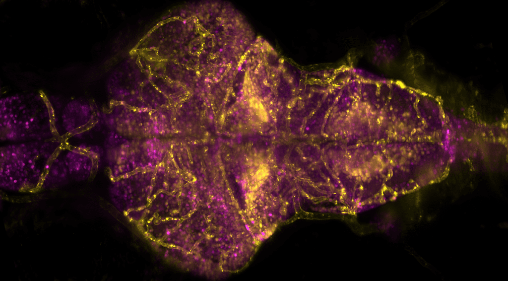
Vascular system and calcium levels in the larval zebrafish brain, recorded by Michael Weber (Morgridge Institute for Research) using a Flamingo T-SPIM light sheet microscope. Specimen kindly provided by Armin Bahl (Engert Lab, Harvard University; now University of Konstanz).
Morgridge investigators Jan Huisken and Kevin Eliceiri will lead an initiative to develop and advance light sheet microscopy technology through a grant funded by the Arnold and Mabel Beckman Foundation.
As one of eight awardees, the team at Morgridge will receive $1.2 million this spring to support their proposed project over five years.
Light sheet microscopy has the ability to image samples over several hours or days from different angles to generate a 3D view of an entire organism. This fast-imaging technique generates a tremendous amount of data quickly with less phototoxic effects on the sample.
“You’re seeing true biology happening in front of your lens,” says Huisken.
Huisken is a founding leader in light sheet microscopy, and he aims to enable more researchers to adopt the cutting-edge technology into their spaces.
“It’s really a new world for some people,” he says. “Suddenly, they’re doing long time lapse experiments. Suddenly they’re looking at three-dimensional tissues instead of single cells on glass slides. So it can be quite transformative for entire labs and entire departments.”
Mary Halloran, professor of integrative biology and neuroscience at the University of Wisconsin – Madison and a collaborating investigator on the grant, sees light sheet microscopy as integral to her research.
“The use of light sheet imaging gives us an unprecedented ability to image neurons developing throughout an entire zebrafish embryo with a remarkable level of resolution and detail,” Halloran says. “This imaging has revealed new information about the molecular mechanisms that regulate the growth of neuronal axons and the formation of neural circuits during embryogenesis.”
While the size and complexity of light sheet microscopy data has prevented wide-spread adoption of the technology, Huisken and Eliceiri plan to use the Beckman Foundation award to make progress toward a “smart microscope” that also can easily be shared with collaborators.