
03 Jan From ‘blobology’ to atomic precision: Wisconsin’s leadership on cryo-EM imaging
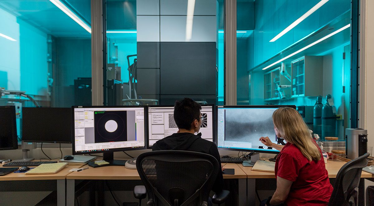
Microscopes have come a long way from what many people remember from their high school biology class. Instead of peering through a lens, scientists can now create 3-D images by shooting beams of electrons at protein structures that have been frozen to hold their shape.
This state-of-the-art technology, called cryo-electron microscopy or cryo-EM, drives a promising collaboration between the UW-Madison Department of Biochemistry and the Morgridge Institute. Recognizing they risked falling behind, the partners worked to build a center that supports a broad range of research on campus and is now a national hub for training and research development.
Cryo-EM is all about getting clearer, more detailed images of the structure of molecules. Scientists need this atomic-level resolution to understand, for example, how proteins malfunction with disease and how to target them when developing a new drug. The specimen is flash-frozen in liquid ethane, which helps reduce the damage that inevitably occurs every time the electrons hit to secure an image.