
15 Feb Morgridge scientist aims to shatter the ‘ballistic barrier’ in imaging
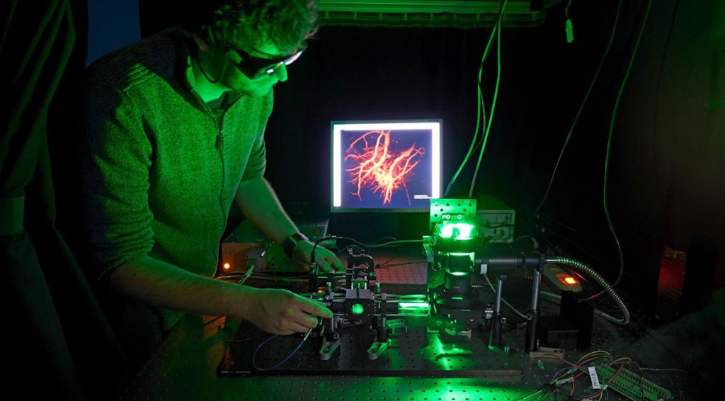
By measuring a nonlinear distortion operator, scientists can image at a higher spatial resolution more easily. Courtesy of Randy Bartels.
Most imaging scientists will tell you that scattered light — rays of light that get rerouted when ping-ponging between obstacles in a frame — is the sworn enemy of clear, crisp biomedical images.
But Randy Bartels envisions a different outcome: By harnessing the chaos of scattered light, it may open up unseen worlds in live biology imaging.
Bartels, a biomedical imaging investigator at the Morgridge Institute for Research, received a Deep Tissue Phase 2 Frontiers of Imaging award on Feb. 13 from the Chan Zuckerberg Initiative (CZI) to develop a new approach to deep tissue imaging. The project addresses a roadblock in biology’s long-sought goal to see inside living tissue to better understand both normal function and disease.
Bartels, also a UW–Madison professor of biomedical engineering, describes the current imaging challenge as the “ballistic barrier.” Normally, researchers want to maximize ballistic light, which is in a straight-line, highly precise trajectory that can penetrate into tissue and provide clear pictures. Think of ballistic light like a bullet slicing through a dense forest and never hitting a tree.
But just as a bullet travelling through a forest will eventually hit a tree, light will inevitably be scrambled by scattering as the light propagates in tissue. Thus, according to Bartels, two problems arise in the imaging process. First, light propagating through tissue is robbed of its precision by all of the “background noise” of scattered light. Second, even when techniques are used to suppress scattered light, the intensity of ballistic light decays exponentially as it moves more deeply into tissue — hence, the ballistic barrier.