
21 Sep New national imaging center has potential to transform medicine
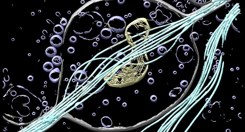
Rendered three-dimensional (3D) view of a neurite from a mouse cortical neuron revealed by cryo-electron tomography (cryo-ET). Microtubules and mitochondria are essential components of cells. In neurons, microtubules are a major structural backbone and are used as transportation highways. Mitochondria provide neurons with energy and metabolites necessary for proper function. Color labels are defined as: membrane (grey), microtubules (cyan), mitochondria (yellow), and vesicles (lavender). UW-Madison collaborative effort between (Joseph Kim) Wright lab and (Tanner Tenpas) Dent lab.
A national research initiative announced today will place the University of Wisconsin-Madison at the forefront of a revolution in imaging fostered by cryo-electron microscopy and cryo-electron tomography — technologies that can illuminate life at the atomic scale.
The National Institutes of Health (NIH) will provide $22.7 million over six years to create a national research and training hub at UW-Madison that will enable scientists across the country to gain access to this game-changing technology.
Cryo-electron microscopy (cryo-EM for short) is a method used to image biological molecules that are flash-frozen to capture them in their native state. No dyes or other alterations are needed to view the structures, which gives scientists a highly accurate picture of true biological function. Scientists can peer into the very surfaces where drugs and proteins interact, where diseases occur and viruses orchestrate their attacks. Cryo-EM has the potential to impact every corner of medicine, from Alzheimer’s disease to vaccine development, protein and cellular engineering and many other areas across all aspects of life sciences research.