
18 Jul Searching for the biomechanical risk factors of preterm birth
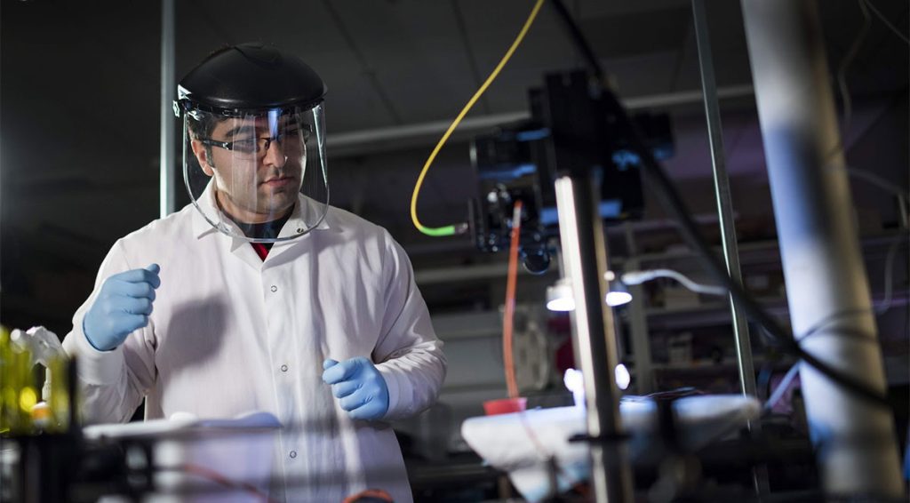
Kayvan Samimi
Preterm birth remains a stubborn and poorly understood medical challenge, but new imaging techniques developed at the Morgridge Institute for Research are helping scientists analyze the key biomechanical features that contribute.
Researchers in the lab of Melissa Skala, a Morgridge biomedical imaging investigator and UW-Madison professor of biomedical engineering, are focusing on fetal membranes — thin tissues that surround the fetus, retain amniotic fluid and protect both the baby and mother from infection. These membranes normally break during labor, but preterm rupture is often one of the key causes of preterm birth.
Fetal membranes are too thin to be clearly detected by common imaging techniques such as ultrasound and magnetic resonance (MRI). The Skala Lab has helped fill this resolution gap with optical coherence tomography (OCT), a non-invasive technique that gathers data in three dimensions to help determine tissue structure.
Kayvan Samimi, an assistant scientist in the Skala Lab, is leading a multi-year project to image fetal membrane samples that have been provided by partner hospitals after births. Meriter Hospital in Madison and Intermountain Healthcare in Provo, Utah, collectively provided more than 60 fetal membrane samples for the study, from both vaginal and C-section births.