
24 May Unique optical imaging approach sheds light on hidden information within cells
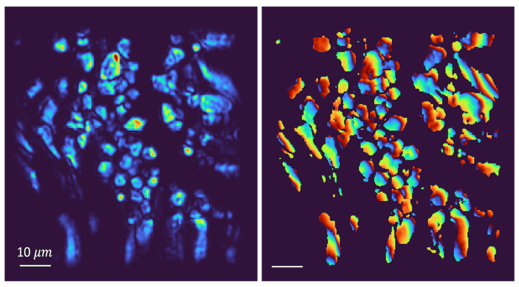
Osteoblasts in developing bone are imaged using third harmonic generation to produce full intensity (left) and full phase (right) images.
Morgridge Investigator Randy Bartels is using optical imaging techniques to answer complex biological questions by observing phenomena that have previously been difficult to capture.
In new research published in the journal Optica, the Bartels Lab and collaborators describe a new approach that looks into biological samples to uncover hidden information within cells and tissues.
The researchers demonstrate for the first time the use of third harmonic generation (THG) holographic microscopy — where the frequency of laser light is tripled when it interacts with materials — to collect measurements of light intensity as well as phase information.
“Third harmonic generation phase has never before been directly measured,” says Yusef Farah, a postdoctoral researcher and first author of the published work. “With this information, we can unlock new material properties or solve problems to generate three dimensional images. This opens up the possibility of new applications in biomedical imaging and material science.”
Light radiates in oscillating waves, forming peaks and valleys that represent the intensity of light. Those waves can phase shift as the light interacts with and reflects off the material through which it is moving.
Bartels explains that most everyone has probably observed this phenomenon without realizing it.
“Caustics in the bottom of a swimming pool — those dancing lines of light moving around,” he says. “When there are ripples at the surface, the light going into the water gets distorted and the phase variation ends up focusing into certain points.”